Brain Injury Screening War Troops Friday, Dec 5 2008 

soldiers1The Institute of Medicine is a government advisory group. This group studies a variety of medical and health issues. They have now issued a new report that urges the US government to screen soldiers who have had traumatic brain injury.

Traumatic brain injury has been a far too common occurrence in the Afghanistan and Iraq wars. Currently as of January and astounding 5,500 US military soldiers have suffered traumatic brain insults that are the result of improvised explosive devices.

The new study has indicated that those soldiers who experience these traumatic brain insults may be more likely to get some long term debilitating conditions. They are at increased risk for having Alzheimer’s dementia, major depression, memory loss and aggression.

The Institute of Medicine has currently recommended that the Department of Defense should devote more money and research to better protecting soldiers against this type of damage. The committee has also suggested that more studies should be performed on the long term side effects of the brain injuries.

The new report by the Institute of Medicine can be found here.

Under a Congressional mandate, the Institute of Medicine (IOM) has reviewed a wide array of biologic, chemical, and physical agents to determine if exposure to the agents might be responsible for the veterans’ long-term health problems. In this 2008 report, Gulf War and Health, Volume 7: Long-term Consequences of Traumatic Brain Injury, the IOM assesses the possible long-term health outcomes of traumatic brain injury (TBI).

The committee details numerous outcomes that might be associated with mild, moderate, and severe injuries. These include: neurologic, endocrine, psychologic outcomes, in addition to problems with social functioning.

In addition to the report’s findings, recommendations are provided for study of blast injuries and the report suggests methods of data-gathering for future research to improve our understanding of blast injuries which ultimately would improve veteran care. The report is part of the IOM’s Gulf War and Health series and was sponsored by the U.S. Department of Veterans Affairs.
Stress from Poverty Causes Brain Functioning Similar to Brain Injury? Tuesday, Dec 2 2008 

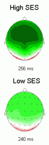
eeg-brainUniversity of California, Berkeley has just come out with new research about the possible effect of poverty on the brain. You can read about the study here. The picture on the left shows the brain functioning in those children who are richer (top) vs. those children who are poorer (bottom).

In a study recently published in the Journal of Cognitive Neuroscience, scientists at UC Berkeley’s Helen Wills Neuroscience Institute and the School of Public Health report that normal 9- and 10-year-olds differing only in socioeconomic status have detectable differences in the response of their prefrontal cortex, the part of the brain that is critical for problem solving and creativity.

Basically the researchers measured the brain’s signals using an EEG brain reader. They found that those who had a lower socioeconomic status were more likely to have poor frontal lobe functioning. The frontal lobes are right behind a person’s forehead and are thought to be involved with attention and planning ahead. Poor functioning in this region of the brain could lead to a variety of symptoms such as attention deficit disorder, apathy, poor insight and poor planning ability. This symptoms fall under the rubric of executive functioning.

The executive system is a theorized cognitive system in psychology that controls and manages other cognitive processes. It is also referred to as the executive function, executive functions, supervisory attentional system, or cognitive control.

The researchers have hypothesized that a poor environment is to blame for these deficits in functioning.

The researchers suspect that stressful environments and cognitive impoverishment are to blame, since in animals, stress and environmental deprivation have been shown to affect the prefrontal cortex. UC Berkeley’s Marian Diamond, professor emeritus of integrative biology, showed nearly 20 years ago in rats that enrichment thickens the cerebral cortex as it improves test performance.

It is still too early to determine if poor frontal lobe functioning is the result of stress from poverty. It could also very well be the reverse, people who have poor frontal lobe functioning may be more likely to be poor. Scientists may need to find ways that improve this type of poor brain functioning. Transcranial magnetic stimulation for instance has been used to improve working memory. So it also might be used to treat a variety of executive function disorders.

Stem Cells to Prevent Brain Damage from Stroke Tuesday, Dec 2 2008 

embryonic-stem-cellThe company Biocompatibles International has just recently completed a new experimental stem cell therapy in a 49 year old man. Stem cells are cells that are located in most organism. Stem cells have the capacity to differentiate themselves into almost any type of cell in the human body. The researchers of this new study have shown that there were no side effects following the surgery using stem cells.

The company is doing a Phase 1/2 trial in 20 different people. The trial is supposed to find out whether the treatment is safe for those who have undergone severe strokes. This specific patient regained some of his ability to speak and his paralysis also disappeared over the course of the treatment. However, the researchers said it was not clear yet whether this improvement was due to the stem cells themselves. The man also had brain surgery to reduce pressure on his brain, which could also have been the cause of the improvement. The researchers stressed that this was just a safety test and not an efficacy test of using stem cells.

People who have brain damage from strokes often have many incapacitating difficulties. Researchers have been investigating ways to treat and prevent stroke damage in addition to stroke rehab. In the past, scientists have been able to transplant brain cells that were created from stem cells into the brain’s of rats. Implanting these new brain cells was able to restore some of the rats functioning after they had an induced stroke. For this specific study, the researchers used the less controversial adult stem cells that they obtained from a healthy donor. The scientists then programmed the stem cells to deliver a protein named GLP-1. The GLP-1 protein is able to protect against brain cell death after a person undergoes a stroke.  So implanting these stem cells in the brain might reduce stroke damage. Approval for this specific therapy may not come until 2012.

Severe Brain Injury Prevention Sciam Tuesday, Dec 2 2008 

brain-injury-treatmentScientific American Mind has a new article about the treatment of brain injuries. They say that there are a variety of new methods of preventing brain damage from getting worse. These methods can be used immediately on a person after they have suffered a traumatic brain insult.  I think in the future we will see better preventative measures for brain injuries and also better treatment as well.

  • Doctors can often save the life of a victim of severe traumatic brain injury if they reach the patient soon enough—but are far less able to preserve the person’s mental abilities.
  • The best hope for improved healing lies neither in new medications, which have been disappointing so far, nor in exotic fixes involving stem cells and neural regeneration, which are at least a decade away.
  • The biggest gains in cognitive recovery will likely result from advances in emergency room and intensive care practices such as slowing the brain’s metabolism by cooling the body, removing part of the skull to relieve intracranial pressure and injecting an experimental polymer “glue” to repair damaged brain cells.

Helmet Research Brain Damage Prevention Monday, Dec 1 2008 

army-helmetAn extremely high number of Iraq and Afghanistan war veterans have sustained a traumatic brain injury or insult. These injuries can cause a number of problems for the individual soldier. Scientists are continually creating novel ways to diagnose and also to treat traumatic brain injuries. However, the ideal thing would be to prevent these head injuries from occurring in the first place.

Now some new research has come out of the Lawrence Livermore National Laboratory. Scientists there have been investigating the mechanics of how a bomb blast is able to affect the functioning of a soldier’s brain. Helmets in current use have been specially designed so as to minimize the amount of force from a blast impact. Even though helmets are protective, scientists have found that some helmet designs have a gap that is located between the helmet and a soldier’s skull. This gap actually has the potential to amplify some of the pressure that is due to a bomb blast. So in the future scientists may need to design helmets that will not do this. This may lead to better helmets that are more likely to prevent brain injuries. This could potentially reduce a large amount of the damage that is the result of brain insults.

The study is named “Skull Flexure from Blast Waves: A New Mechanism for Brain Injury with Implications for Helmet Design”:

(ii) Instead, a blast wave causes the skull to dynamically flex inwards and ripple outwards, creating localized regions of high and low pressure that produce large pressure gradients that are swept through the brain. The loads associated with even small amounts of skull flexure from a non-lethal blast wave are comparable to or greater than those in a typical injury-inducing impact, and should not be neglected.

(iii) Helmets can affect the interaction of the blast with the head in unexpected ways. In the absence of padding, the existence of a clearance gap between the helmet and the head allows underwash of the blast wave to create amplified pressures that act directly on the skull. Padding inhibits this underwash, but more strongly couples the helmet motion to the head, which can increase head accelerations and transmit blast-induced flexure to the skull.