Uncategorized
johnw456
12:43 am
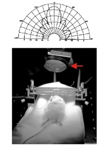 Researchers from Arizona State University are investigating the use of low-frequency and also low-power ultrasonic pulses on the brain. The researchers believe that ultrasound can manipulate the functioning of the brain. The low frequency ultrasound has the capability of non-invasively penetrating a person’s skull. It can then touch practically any area of the brain. The ultrasound can then modulate neural activity by altering the activity of voltage-gated ion channels. Doing this allows scientists to stimulate specific areas of the brain with relative selectivity.
The researchers believe that ultrasonic neuromodulation may eventually become a treatment modality for all sorts of brain disorders. It could treat parkinson’s, autism, epilepsy, mental retardation and many other things.
Ultrasound can also be used to lower the metabolic rate of the entire brain. Doing this would allow a reduction in the amount of damage that occurs after undergoing a traumatic brain insult. Often when their is a trauma to the head, there is a lot of secondary damage that may not occur until minutes to hours after the insult. The ultrasound, in theory, could reduce the metabolism of the brain. This could lead to a reduction in the amount of chemical reactions that lead to neuron brain cell death.
One of the researchers has said that a helmet could have an array of ultrasound transducers on it. When the helmet detected a traumatic brain insult it could be turned on automatically. This would enable the array to modulate various neuroprotective mechanisms in the brain. It would allow a reduction in the amount of brain damage that person gets.
Leave a Response »
Uncategorized
brain damage rehabilitation, Brain disorders, brain injury, brain injury rehab, brain injury rehabilitation, fMRI, functional MRI, Radiological Society of North America, robotic rehab tool, Stroke, stroke rehab, stroke rehabilitation, traumatic brain damage, traumatic brain damage rehab, traumatic brain injury, traumatic brain injury rehab johnw456
10:16 pm
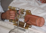 Researchers have recently used a custom hand exercise robotic machine and fMRI brain imaging to help chronic stroke patients restore some functioning. The robotic machine was used for doing several rehabilitation exercises. The doctors monitored the changes in the fMRI as the patients regained some of their previous functioning. This is basically using the brain’s neuroplastic mechanisms to restore some previous functioning. Here is the press release.
CHICAGO — Research scientists using a novel, hand-operated robotic device and functional MRI (fMRI) have found that chronic stroke patients can be rehabilitated, according to a study presented today at the annual meeting of the Radiological Society of North America (RSNA). This is the first study using fMRI to map the brain in order to track stroke rehabilitation.
“We have shown that the brain has the ability to regain function through rehabilitative exercises following a stroke,” said A. Aria Tzika, Ph.D., director of the NMR Surgical Laboratory at Massachusetts General Hospital (MGH) and Shriners Burn Institute and assistant professor in the Department of Surgery at Harvard Medical School in Boston. “We have learned that the brain is malleable, even six months or more after a stroke, which is a longer period of time than previously thought.”
Post-stroke rehabilitation is nothing new and has already been reported by researchers in the past. See here and also here and here. So this is not the first time that fMRI has been used to track progress among stroke patients and the press release may be wrong. It has also been known for some time that the brain’s neuroplasticity allows for improved functioning that can be quite long lasting. However, this may be the first use of a robotic device such as the one used.

Figure 16. This fMRI image illustrates the area in the brain that corresponds with hand use of a patient before training (left), after eight weeks of training (middle), and one month after training was completed (right) at a 60percent effort level.
In the future researchers may use real time brain imaging to shape the brain among those who are brain injured. It would allow real-time feedback. A person would be able to look at their own brain scan and attempt to change their own brain waves by themselves. It could potentially shape the brain for beneficial effect among those who currently have poor functioning as a result of brain injuries.
Leave a Response »
Uncategorized
Brain Damage, Brain Damage Prevention, Brain Damage Treatment, brain damage war, Brain disorders, Brain Imaging, brain injury, brain injury dementia, Brain Injury Prevention, brain injury treatment, brain injury war, brain trauma dementia, brain trauma war, mental health issues, traumatic brain damage, Traumatic brain damage prevention, Traumatic Brain Damage Treatment, traumatic brain injury, Traumatic brain injury prevention, Traumatic Brain Injury Treatment, war wounds johnw456
4:00 am
 The Institute of Medicine is a government advisory group. This group studies a variety of medical and health issues. They have now issued a new report that urges the US government to screen soldiers who have had traumatic brain injury.
Traumatic brain injury has been a far too common occurrence in the Afghanistan and Iraq wars. Currently as of January and astounding 5,500 US military soldiers have suffered traumatic brain insults that are the result of improvised explosive devices.
The new study has indicated that those soldiers who experience these traumatic brain insults may be more likely to get some long term debilitating conditions. They are at increased risk for having Alzheimer’s dementia, major depression, memory loss and aggression.
The Institute of Medicine has currently recommended that the Department of Defense should devote more money and research to better protecting soldiers against this type of damage. The committee has also suggested that more studies should be performed on the long term side effects of the brain injuries.
The new report by the Institute of Medicine can be found here.
Under a Congressional mandate, the Institute of Medicine (IOM) has reviewed a wide array of biologic, chemical, and physical agents to determine if exposure to the agents might be responsible for the veterans’ long-term health problems. In this 2008 report, Gulf War and Health, Volume 7: Long-term Consequences of Traumatic Brain Injury, the IOM assesses the possible long-term health outcomes of traumatic brain injury (TBI).
The committee details numerous outcomes that might be associated with mild, moderate, and severe injuries. These include: neurologic, endocrine, psychologic outcomes, in addition to problems with social functioning.
In addition to the report’s findings, recommendations are provided for study of blast injuries and the report suggests methods of data-gathering for future research to improve our understanding of blast injuries which ultimately would improve veteran care. The report is part of the IOM’s Gulf War and Health series and was sponsored by the U.S. Department of Veterans Affairs.
Leave a Response »
neuromodulation
brain alteration, Brain Damage, Brain Damage Treatment, Brain disorders, brain injuries, brain injury, brain injury treatment, Brain manipulation, International Neuromodulation Society, neuromodulation, neuromodulation techniques, neuroscientist, traumatic brain damage, Traumatic Brain Damage Treatment, traumatic brain injury, Traumatic Brain Injury Treatment johnw456
6:09 pm
 The International Neuromodulation Society (INS) is currently assembling a meeting from today until december 7th. They will have poster presentations and scientific oral presentations. The scientists going to this meeting will talk about a variety of neuromodulation techniques that may be able to treat many brain based disorders. Future ways of neuromodulation may be able to treat brain injuries as well. You can read about the gathering at this press release. Here are some excerpts from the press release.
Stroke Rehabilitation. The idea that the adult brain can reorganize to restore normal function after sustaining trauma is well accepted, but the means to achieving such rehabilitation is still a work-in-progress. A recent clinical trial – EVEREST – tested the idea that directly stimulating the brain with electrical signals coupled with classical rehabilitation techniques may speed the recovery of movement. Dr. Robert Levy, a neurosurgeon at Northwestern University in Chicago,will report new analyses of the trial data which suggest that this approach may be successful in some, but not all, stroke victims.
New Technologies. A special session on novel neuromodulation therapies will explore the cutting edge of electrical and optical technologies for studying and altering pathological brain activity. These include advances in the use of light as a tool to alter neural activity, new uses of transcranial magnetic stimulation and the use of oscillating electric fields to regrow spinal nerves after injury. Dr Karl Deisseroth, a neuroscientist at Stanford University, will deliver the keynote address, reporting on his work to engineer light-reactive molecules into neural circuits to manipulate and study patterns of activity in model organisms.
Leave a Response »
virtual reality
acquired brain injury, brain damage therapy, Brain Damage Treatment, brain injury, brain injury therapy, Neuron, neuroplasticity, virtual reality, virtual reality therapy johnw456
12:11 am
 Researchers are now doing a new virtual reality rehabilitation program for those with cognitive deficits that are due to acquired brain damage. Virtual reality allows researchers to control the sensory inputs that go into a person’s brain. This type of therapy may be able to improve many cognitive symptoms associated with brain injuries such as anoxic brain injury, hypoxic brain injury and brain stem injury. In the future virtual reality may become an extremely useful tool that researchers will increasingly use to shape the brain. You can watch the video about this treatment here. The press release can be found here. This is certainly a fascinating development that should help many who currently have many devastating brain injuries. This type of brain manipulation method will likely be much safer than other brain manipulation tools such as transcranial direct current stimulation and transcranial magnetic stimulation.
The PREVIRNEC platform enables therapists to personalize treatment plans: intensive rehabilitation can be programmed automatically for the required length of time, the results monitored and the level of difficulty adjusted according to patients’ performance in previous sessions.
A multidisciplinary team of researchers is currently working on the project. The UPC’s CREB, coordinated by lecturer Daniela Tost, is in charge of 3D software, the Guttmann Institute, a benchmark in neurorehabilitation, contributes neuropsychological and therapeutic knowledge, and a group from the Rovira i Virgili University is responsible for distributed software.
The aim of this project is to use software to meet the treatment needs of patients with acquired brain damage. The software promotes the rehabilitation of affected cognitive functions by representing everyday, real life situations in a virtual world.
Leave a Response »
Uncategorized
Brain Damage, brain damage stimulation, brain injury, brain injury stimulation, Brain Stimulation, deep brain stimulation, health, medicine, neuromodulation, neuron stimulation, Neurotechnology, news, non-invasive brain stimulation, transcranial magnetic stimulation, traumatic brain damage, Traumatic Brain Damage Treatment, traumatic brain injury, Traumatic Brain Injury Treatment, Ultrasound, Ultrasound Neuromodulation, vagal nerve stimulation johnw456
11:00 pm
 I’ve mentioned in the past about scientists using transcranial magnetic stimulation and transcranial direct current stimulation as methods to improve brain injury associated with stroke. These are non-invasive brain stimulation techniques that could improve a variety of brain disorders and brain injuries. Recently, for instance, transcranial magnetic stimulation has been used to awaken a coma victim.
At first, there was little change in Villa’s condition, but after around 15 sessions something happened. “You started talking to him and he would turn his head and look at you,” says McAndrews. “That was huge.”
So these are very powerful and selective tools that can do all sorts of interesting things. They can selectively alter the functioning of the brain and have relatively few side effects associated with there use.
What about the future of brain manipulation? Recently, scientists have used ultrasound to modify the functioning of the brain.
In a twist on nontraditional uses of ultrasound, a group of neuroscientists at Arizona State University has developed pulsed ultrasound techniques that can remotely stimulate brain circuit activity. Their findings, published in the Oct. 29 issue of the journal Public Library of Science (PLoS) One, provide insights into how low-power ultrasound can be harnessed for the noninvasive neurostimulation of brain circuits and offers the potential for new treatments of brain disorders and disease.
Ultrasound is has a much better targeting accuracy than transcranial magnetic stimulation. It can selectively excite brain regions that are 1 mm in size while transcranial magnetic stimulation only has a focal range of 1 cm or greater. Ultrasound can also non-invasively stimulate almost any brain region. Whereas current transcranial magnetic stimulation is limitied to 1-2 cm from a person’s skull.
US may be able to confer a spatial resolution similar to those achieved by currently implemented neuromodulation strategies such as vagal nerve stimulation and DBS, which have been shown to possess high therapeutic value
With this type of neuromodulation power scientists may be able to excite or inhibit any brain regions. This would allow a selective way of altering the brain through neuroplastic mechanisms.
Leave a Response »
Poverty
Brain Damage, Brain Damage Prevention, brain injury, Brain Injury Prevention, cognitive neuroscience, executive functioning, poor, Poverty, Poverty Brain Functioning, rich, socioeconomic status, stress, transcranial magnetic stimulation, Traumatic brain damage prevention johnw456
10:04 pm
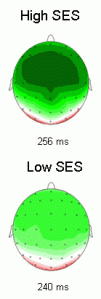 University of California, Berkeley has just come out with new research about the possible effect of poverty on the brain. You can read about the study here. The picture on the left shows the brain functioning in those children who are richer (top) vs. those children who are poorer (bottom).
In a study recently published in the Journal of Cognitive Neuroscience, scientists at UC Berkeley’s Helen Wills Neuroscience Institute and the School of Public Health report that normal 9- and 10-year-olds differing only in socioeconomic status have detectable differences in the response of their prefrontal cortex, the part of the brain that is critical for problem solving and creativity.
Basically the researchers measured the brain’s signals using an EEG brain reader. They found that those who had a lower socioeconomic status were more likely to have poor frontal lobe functioning. The frontal lobes are right behind a person’s forehead and are thought to be involved with attention and planning ahead. Poor functioning in this region of the brain could lead to a variety of symptoms such as attention deficit disorder, apathy, poor insight and poor planning ability. This symptoms fall under the rubric of executive functioning.
The executive system is a theorized cognitive system in psychology that controls and manages other cognitive processes. It is also referred to as the executive function, executive functions, supervisory attentional system, or cognitive control.
The researchers have hypothesized that a poor environment is to blame for these deficits in functioning.
The researchers suspect that stressful environments and cognitive impoverishment are to blame, since in animals, stress and environmental deprivation have been shown to affect the prefrontal cortex. UC Berkeley’s Marian Diamond, professor emeritus of integrative biology, showed nearly 20 years ago in rats that enrichment thickens the cerebral cortex as it improves test performance.
It is still too early to determine if poor frontal lobe functioning is the result of stress from poverty. It could also very well be the reverse, people who have poor frontal lobe functioning may be more likely to be poor. Scientists may need to find ways that improve this type of poor brain functioning. Transcranial magnetic stimulation for instance has been used to improve working memory. So it also might be used to treat a variety of executive function disorders.
One Response »
stem cells
Brain Damage, Brain Damage Prevention, Brain Damage Treatment, brain injury, Brain Injury Prevention, brain injury treatment, Neuron, Neuron replacement, neurons, Neurotechnology, Stem Cell, Stem Cell Research, Stem Cell Therapy, stem cells, traumatic brain damage, Traumatic brain damage prevention, Traumatic Brain Damage Treatment, traumatic brain injury, Traumatic brain injury prevention, Traumatic Brain Injury Treatment johnw456
9:09 pm
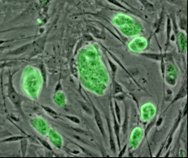 The company Biocompatibles International has just recently completed a new experimental stem cell therapy in a 49 year old man. Stem cells are cells that are located in most organism. Stem cells have the capacity to differentiate themselves into almost any type of cell in the human body. The researchers of this new study have shown that there were no side effects following the surgery using stem cells.
The company is doing a Phase 1/2 trial in 20 different people. The trial is supposed to find out whether the treatment is safe for those who have undergone severe strokes. This specific patient regained some of his ability to speak and his paralysis also disappeared over the course of the treatment. However, the researchers said it was not clear yet whether this improvement was due to the stem cells themselves. The man also had brain surgery to reduce pressure on his brain, which could also have been the cause of the improvement. The researchers stressed that this was just a safety test and not an efficacy test of using stem cells.
People who have brain damage from strokes often have many incapacitating difficulties. Researchers have been investigating ways to treat and prevent stroke damage in addition to stroke rehab. In the past, scientists have been able to transplant brain cells that were created from stem cells into the brain’s of rats. Implanting these new brain cells was able to restore some of the rats functioning after they had an induced stroke. For this specific study, the researchers used the less controversial adult stem cells that they obtained from a healthy donor. The scientists then programmed the stem cells to deliver a protein named GLP-1. The GLP-1 protein is able to protect against brain cell death after a person undergoes a stroke. So implanting these stem cells in the brain might reduce stroke damage. Approval for this specific therapy may not come until 2012.
Leave a Response »
Uncategorized
Brain Damage, Brain Damage Prevention, Brain Damage Treatment, brain injury, Brain Injury Prevention, brain injury treatment, health, medicine, Neurotechnology, traumatic brain damage, Traumatic brain damage prevention, Traumatic Brain Damage Treatment, traumatic brain injury, Traumatic brain injury prevention, Traumatic Brain Injury Treatment johnw456
8:44 pm
 Scientific American Mind has a new article about the treatment of brain injuries. They say that there are a variety of new methods of preventing brain damage from getting worse. These methods can be used immediately on a person after they have suffered a traumatic brain insult. I think in the future we will see better preventative measures for brain injuries and also better treatment as well.
- Doctors can often save the life of a victim of severe traumatic brain injury if they reach the patient soon enough—but are far less able to preserve the person’s mental abilities.
- The best hope for improved healing lies neither in new medications, which have been disappointing so far, nor in exotic fixes involving stem cells and neural regeneration, which are at least a decade away.
- The biggest gains in cognitive recovery will likely result from advances in emergency room and intensive care practices such as slowing the brain’s metabolism by cooling the body, removing part of the skull to relieve intracranial pressure and injecting an experimental polymer “glue” to repair damaged brain cells.
Leave a Response »
Brain Damage Treatment and Neurotechnology and Transcranial Direct Current Stimulation and Uncategorized
Neurotech, Neurotechnology, Stroke, Transcranial Direct Current Stimulation, Traumatic Brain Damage Treatment, Traumatic Brain Injury Treatment johnw456
4:06 am
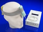 Transcranial direct current stimulation is a way of non-invasively stimulating the brain with a small amount of electricity. Basically you attach two electrodes to your head and run a 1 to 2 milliamp current through them. This amount electricity is much less than the more drastic electroshock and can be performed on yourself while you are fully awake. A man demonstrates how the technique works in this youtube video. Transcranial direct current stimulation can excite or inhibit brain regions that are close to a person’s skull. It cannot penetrate very far into the brain, so it is limited in its targeting ability. Transcranial direct current stimulation may be used for people who have sustained traumatic brain injuries including those who have had a stroke. Here is a paper discussing this new treatment method for stroke patients.
Electrical brain stimulation, a technique developed many decades ago and then largely forgotten, has re-emerged recently as a promising tool for experimental neuroscientists, clinical neurologists and psychiatrists in their quest to causally probe cortical representations of sensorimotor and cognitive functions and to facilitate the treatment of various neuropsychiatric disorders. In this regard, a better understanding of adaptive and maladaptive plasticity in natural stroke recovery over the last decade and the idea that brain polarization may modulate neuroplasticity has led to the use of transcranial direct current stimulation (tDCS) as a potential enhancer of natural stroke recovery. We will review tDCS’s successful utilization in pilot and proof-of-principle stroke recovery studies, the different modes of tDCS currently in use, and the potential mechanisms underlying the neural effects of tDCS.
One Response »
Next Page »
 Researchers from Arizona State University are investigating the use of low-frequency and also low-power ultrasonic pulses on the brain. The researchers believe that ultrasound can manipulate the functioning of the brain. The low frequency ultrasound has the capability of non-invasively penetrating a person’s skull. It can then touch practically any area of the brain. The ultrasound can then modulate neural activity by altering the activity of voltage-gated ion channels. Doing this allows scientists to stimulate specific areas of the brain with relative selectivity.
 Researchers have recently used a custom hand exercise robotic machine and fMRI brain imaging to help chronic stroke patients restore some functioning. The robotic machine was used for doing several rehabilitation exercises. The doctors monitored the changes in the fMRI as the patients regained some of their previous functioning. This is basically using the brain’s neuroplastic mechanisms to restore some previous functioning. Here is the
 The Institute of Medicine is a government advisory group. This group studies a variety of medical and health issues. They have now issued a new report that urges the US government to screen soldiers who have had


